Bone age from a hand-wrist radiograph in seconds
Upload the radiograph and select the patient's age group. The matching specialist model estimates bone age and sex; the result is ready as a printable report for physician review.
- First analysis is free
- JPG, PNG and DICOM
- KVKK-aligned, encrypted storage
Clinical decision support only. Results must be reviewed by a qualified healthcare professional.
Try it without signing up
Real model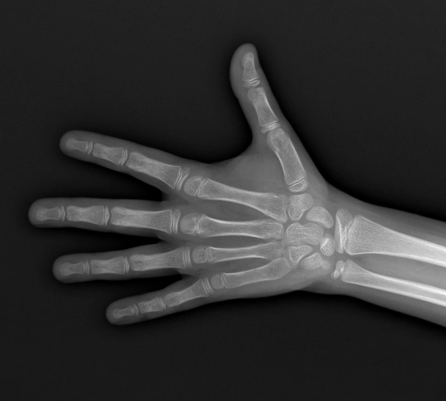
De-identified sample image. The patient's verified age and sex are known, so you can check the model against ground truth. It does not use your free trial.
Specialist models per age group
Results in months under 18 and in years over 18. You pick the group; if you don't know, the system decides from the image.
Printable clinical report
Save to PDF with report number, institution details and a physician signature line. Enter the chronological age and the deviation is computed for you.
Encrypted storage, KVKK-aligned
Images and results are stored encrypted. You can access your own history and request deletion at any time.
Likely age range
Alongside the single value, a range derived from the model's age distribution is shown (e.g. 147 months, likely range 138–156). The clinical margin of error is 7–11 months depending on the age band.
Heatmap and review screen
A Grad-CAM heatmap shows where the model looked most when deciding. Zoom up to 6×, pan and measure between two points; measurements are added to the report.
Longitudinal follow-up
Pick two analyses from your history: change in bone age, growth rate and a projected time to maturity on one screen. Linear assumption; physician judgement prevails.
Everything in the 10 September 2026 release is on the What's New page.
Workflow
- 1Create a free account and log in; your first analysis is on us.
- 2Upload a complete hand-wrist radiograph (JPG, PNG or DICOM).
- 3Select whether the patient is under or over 18 — or leave it to the system; the specialist model estimates bone age and sex.
- 4Review the result, compare with chronological age and print the report.
Clinical Limits
- Not a standalone diagnosis.
- Requires physician or radiology specialist review.
- Image quality, positioning, artifacts, and atypical anatomy may affect the result.
Validation results and model details are on the methodology page.
Create a free account — your first analysis is on us
Looking for an annual licence for your institution? Request a quote